Welcome to the Amira-Avizo Software Use Case Gallery
Below you will find a collection of use cases of our 3D data visualization and analysis software. These use cases include scientific publications, articles, papers, posters, presentations or even videos that show how Amira-Avizo Software is used to address various scientific and industrial research topics.
Use the Domain selector to filter by main application area, and use the Search box to enter keywords related to specific topics you are interested in.

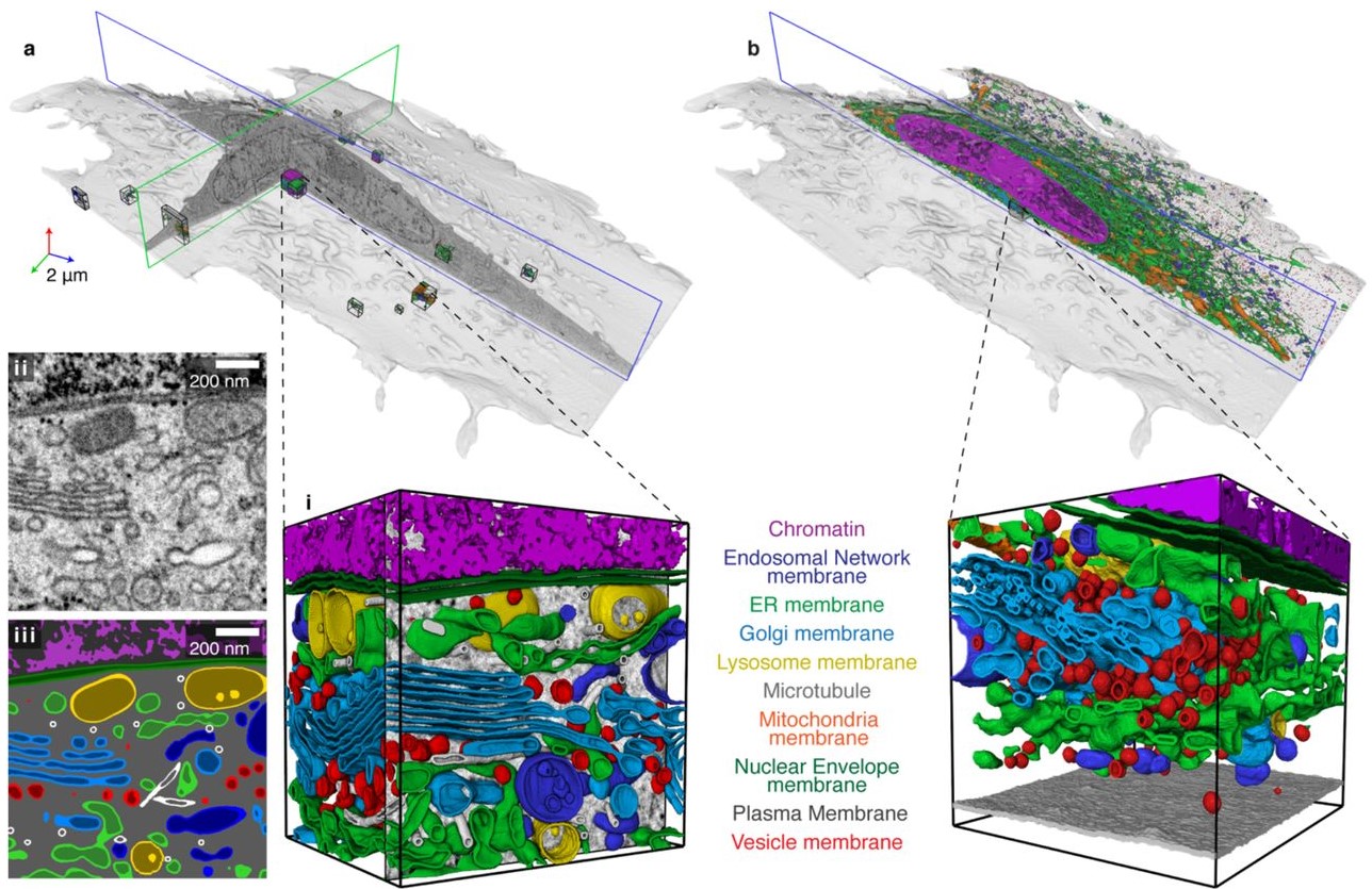
Protocols for Generating Surfaces and Measuring 3D Organelle Morphology Using Amira
High-resolution 3D images of organelles are of paramount importance in cellular biology. Although light microscopy and transmission electron microscopy (TEM) have provided the standard for imaging cellular structures, they cannot provide 3D images.
However, recent technological advances such as serial block-face scanning electron microscopy (SBF-SEM) and focused ion beam scanning electron microscopy (FIB-SEM) provide the tools to create 3D images for the ultrastructural analysis of org... Read more
Edgar Garza-Lopez, Zer Vue, Prasanna Katti, Kit Neikirk, Michelle Biete, Jacob Lam, Heather K. Beasley, Andrea G. Marshall, Taylor A. Rodman, Trace A. Christensen, Jeffrey L. Salisbury, Larry Vang, Margaret Mungai, Salma Ash Shareef, Sandra A. Murray, Jianqiang Shao, Jennifer Streeter, Brian Glancy, Renata O. Pereira1, E. Dale Abel, and Antentor Hinton, Jr.
Automatic whole cell organelle segmentation in volumetric electron microscopy
Cells contain hundreds of different organelle and macromolecular assemblies intricately organized relative to each other to meet any cellular demands. Obtaining a complete understanding of their organization is challenging and requires nanometer-level, three-dimensional reconstruction of whole cells. Even then, the immense size of datasets and large number of structures to be characterized requires generalizable, automatic methods.
To meet this challenge, we developed an analy... Read more
Larissa Heinrich, Davis Bennett, David Ackerman, Woohyun Park, John Bogovic, View ORCID ProfileNils Eckstein, Alyson Petruncio, Jody Clements, C. Shan Xu, Jan Funke, Wyatt Korff, Harald F. Hess, Jennifer Lippincott-Schwartz, Stephan Saalfeld, Aubrey V. Weigel, COSEM Project Team
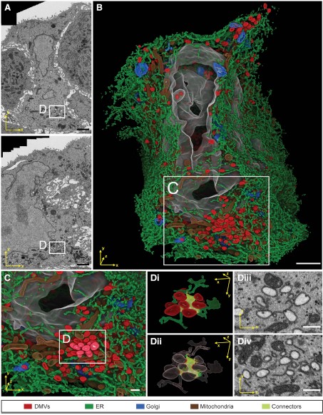
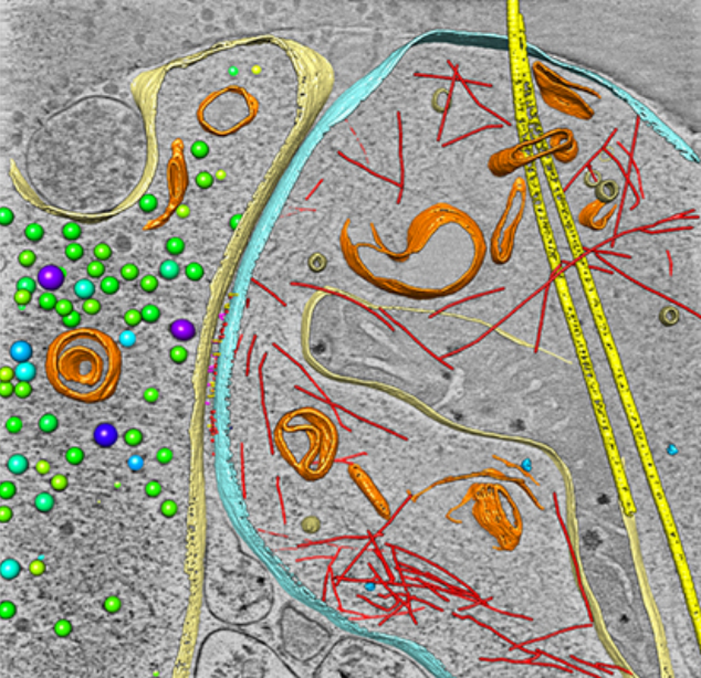
Integrative Imaging Reveals SARS-CoV-2-Induced Reshaping of Subcellular Morphologies
Cortese et al. use integrative imaging techniques to generate a publicly available repository of morphological alterations induced by SARS-CoV-2 in lung cells. Accumulation of ER-derived double-membrane vesicles, the viral replication organelle, occurs concomitantly with cytoskeleton remodeling and Golgi fragmentation. Pharmacological alteration of cytoskeleton dynamics restricts viral replication and spread.
Pathogenesis induced by SARS-CoV-2 is thought to result from both an inflamma... Read more
Mirko Cortese, Ji-Young Lee, Berati Cerikan, ..., Laurent Chatel-Chaix, Yannick Schwab, Ralf Bartenschlager
Membrane architecture of pulmonary lamellar bodies revealed by post-correlation on-lamella cryo-CLEM
Lamellar bodies (LBs) are surfactant rich organelles in alveolar type 2 cells. LBs disassemble into a lipid-protein network that reduces surface tension and facilitates gas exchange at the air-water interface in the alveolar cavity. Current knowledge of LB architecture is predominantly based on electron microscopy studies using disruptive sample preparation methods. We established a post-correlation on-lamella cryo-correlative light and electron microscopy approach for cryo-FIB milled lung ce... Read more
Steffen Klein, Benedikt H. Wimmer, Sophie L. Winter, Androniki Kolovou, Vibor Laketa, Petr Chlanda
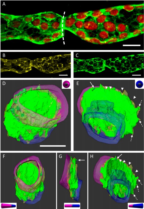
3D Dissection of Structural Membrane-Wall Contacts in Filamentous Moss Protonemata
Cell-to-cell contact is essential for communication and development of multicellular organisms. A prerequisite is the passage through membranes. That way, molecular exchange and information flow is regulated via hormones, membrane proteins and pores.
In plants, the rigid cell walls prevent large membrane contact areas between protoplasts. Only plasmodesmata, minute channels between adjacent cells, form direct connections. Often, molecular data of the proteins involved are manifold but t... Read more
Dominik Harant and Ingeborg Lang
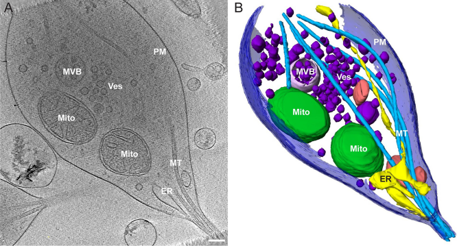
Morphology of mitochondria in spatially restricted axons revealed by cryo-electron tomography
Neurons project axons to local and distal sites and can display heterogeneous morphologies with limited physical dimensions that may influence the structure of large organelles such as mitochondria. Using cryo-electron tomography (cryo-ET), we characterized native environments within axons and presynaptic varicosities to examine whether spatial restrictions within these compartments influence the morphology of mitochondria. Segmented tomographic reconstructions revealed distinctive morphologi... Read more
Tara D. Fischer, Pramod K. Dash, Jun Liu, M. Neal Waxham
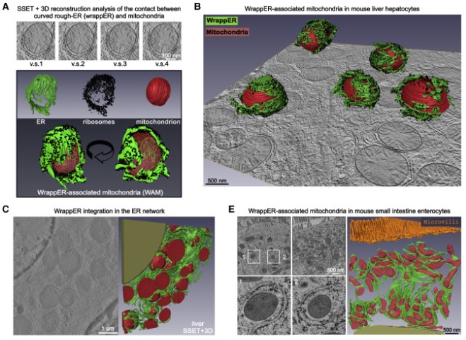
Mitochondria-rough-ER contacts in the liver regulate systemic lipid homeostasis
In this work, we studied mitochondria-rER contacts in vivo by serial section electron tomography (SSET) and 3D reconstruction analysis of cryo-fixed mouse tissue samples. We characterized this inter-organelle association as mitochondria tightly wrapped by sheets of curved rER (wrappER). Further, we used multi-omics and genetic approaches to obtain evidence that the wrappER is a distinct intracellular compartment and demonstrate the importance of wrappER-mitochondria contacts for v... Read more
Irene Anastasia, Nicolò Ilacqua, Andrea Raimondi, Philippe Lemieux, Rana Ghandehari-Alavijeh, Guilhem Faure, Sergei L. Mekhedov, Kevin J. Williams, Federico Caicci, Giorgio Valle, Marta Giacomello, Ariel D. Quiroga, Richard Lehner, Michael J. Miksis, Katalin Toth, Thomas Q. de Aguiar Vallim, Eugene V. Koonin, Luca Scorrano, Luca Pellegrini
As key functional units in neural circuits, different types of neuronal synapses play distinct roles in brain information processing, learning, and memory. Synaptic abnormalities are believed to underlie various neurological and psychiatric disorders. Here, by combining cryo-electron tomography and cryo-correlative light and electron microscopy, we distinguished intact excitatory and inhibitory synapses of cultured hippocampal neurons, and visualized the in situ 3D organization of ... Read more
Chang-Lu Tao, Yun-Tao Liu, Rong Sun, Bin Zhang, Lei Qi, Sakar Shivakoti, Chong-Li Tian, Peijun Zhang, Pak-Ming Lau, Z. Hong Zhou and Guo-Qiang Bi
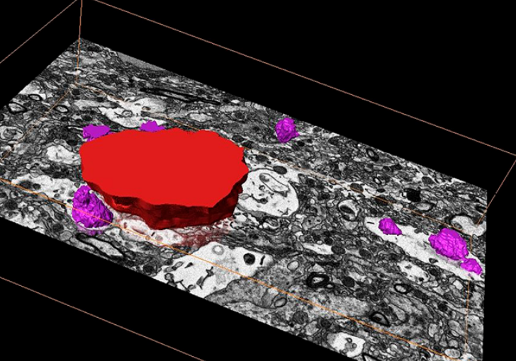
Corpora amylacea are cell-derived structures that appear physiologically in the aged human brain. While their histological identification is straightforward, their ultrastructural composition and microenvironment at the nanoscale have remained unclear so far, as has their relevance to aging and certain disease states that involve the sequestration of toxic cellular metabolites. Here, we apply correlative serial block-face scanning electron microscopy and transmission electron tomograp... Read more
Paula P. Navarro, Christel Genoud, Daniel Castaño-Díez, Alexandra Graff-Meyer, Amanda J. Lewis, Yvonne de Gier, Matthias E. Lauer, Markus Britschgi, Bernd Bohrmann, Stephan Frank, Jürgen Hench, Gabriel Schweighauser, Annemieke J. M. Rozemuller, Wilma D. J. van de Berg, Henning Stahlberg & Sarah H. Shahmoradian
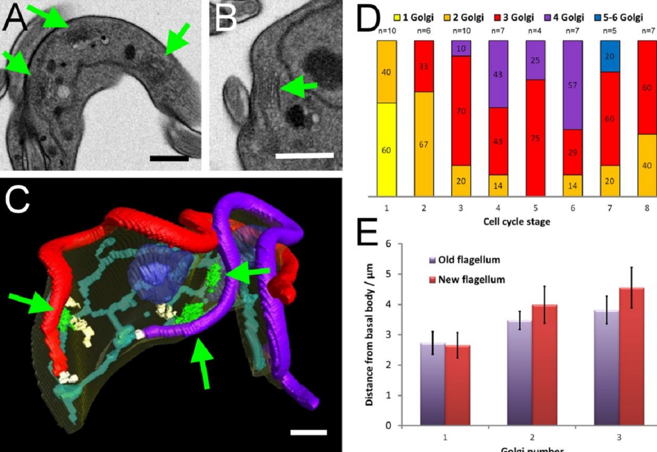
The major mammalian bloodstream form of the African sleeping sickness parasite Trypanosoma bruceimultiplies rapidly, and it is important to understand how these cells divide. Organelle inheritance involves complex spatiotemporal re-arrangements to ensure correct distribution to daughter cells…
Read more
Louise Hughes, Samantha Borrett, Katie Towers, Tobias Starborg, Sue Vaughan
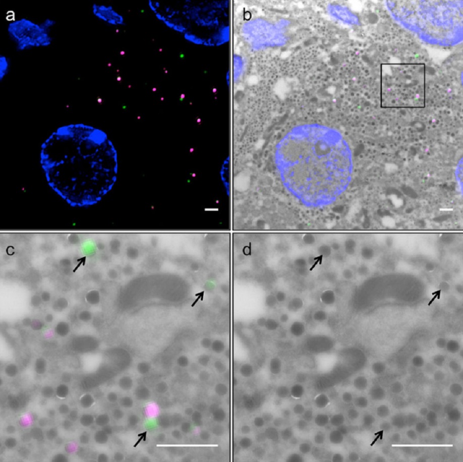
Correlative light and electron microscopy (CLEM) is a powerful approach to investigate the molecular ultrastructure of labeled cell compartments. However, quantitative CLEM studies are rare, mainly due to small sample sizes and the sensitivity of fluorescent proteins to strong fixatives and contrasting reagents for EM. Here, we show that fusion of…
Read more
Andreas Müller, Martin Neukam, Anna Ivanova, Anke Sönmez, Carla Münster, Susanne Kretschmar, Yannis Kalaidzidis, Thomas Kurth, Jean-Marc Verbavatz & Michele Solimena